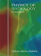
Physics of Radiology, Second Edition
Author: Anthony B. WolbarstISBN: 9781930524224 ISBN10: 1930524226
Published: 2005 | 660 pp. | Hardcover
Price: $ 110.00
SCOPE | March 2007
Dr Wolbarst’s
updated and expanded 660-page book, with 21 additional co-authors, provides a
good grounding in the main areas of medical imaging, both ionising
and non-ionising. This book is intended primarily for
medical radiology residents in the
The book is divided into four
major sections, which are then subdivided into different imaging modalities,
resulting in a total of 60 ‘concise’ chapters! The introduction commences with
a short historical note on medical imaging, the gold standard image quality
parameters, and an overview of film radiography. The first major section,
‘Scientific and Technical Basis’, provides prerequisite learning material for
understanding the core chapters and covers the fundamental basis of ultrasound,
MR and gamma ray imaging. It delves deeper into the physics of X-ray imaging,
and introduces radiation dose, detection and quantification of ionising radiation. The author then goes on to describe the
objective and subjective image quality and system parameters, e.g. ROC and MTF
curves and Poisson statistics, and does this fairly well. PACS is introduced
with DICOM and CADD, which augments the basic chapter on digital image
processing.
The next sections cover the
analogue and digital aspects of diagnostic radiology, together with other imaging
modalities: CT, MRI, nuclear medicine and medical ultrasound. These are
described in depth, covering physics, instrumentation, clinical and safety
aspects. However, readers should note that the standards covered are US only.
One short chapter is solely devoted to describing the evolving and experimental
technologies in medical imaging, including terahertz, microwave, thermography and nanotechnology.
The final two sections cover
‘Radiation Dose, Biological Effects, Risk, and Radiation Safety’ and ‘Radiation
Safety and Emergency Response’. It considers in great detail the dosimetric, stochastic, deterministic and safety aspects of
ionising radiation, and also ways to respond to major
radiological and nuclear emergencies with reference to past incidents.
The book is written in a
clear, relaxed and straightforward style. It is very descriptive and well
laid-out. The author provides numerous ‘real-life’ analogies prior to
explaining the related physics with exercises, and actually does it quite well.
However, the text at times seemed convoluted in explaining the physical
processes, and there was a bit too much description. Nonetheless, the book is
up-to-date, with many references, and can be used as a handbook.
There are minor discrepancies
in the book, and grammatical errors. Moreover, there is a slight overlap
between chapters, although this is useful as an aide-memoire.
Also, there are plenty of illustrations to accompany the text, and each does
paint a thousand words!
The book is, in my opinion, a
very good text for the prospective readers, with a good price tag for a
hardback. I would recommend this book to a department library and generally to
medical specialists interested in the science and technology of medical
imaging.
Reviewed by
Poole General Hospital NHS
Trust, UK
